EQUIPMENT
At Ophtalmo Vétérinaire, we use the most up-to-date technology to provide your animal with efficient, reliant and secure treatments. Our high performing equipment allows us to diagnose and treat all the ocular conditions of the eye and its adnexa.
EQUIPMENT FOR THE EYE EXAMINATIONS
The eye exam requires specialised equipment such as :
- Slipt lamp to examine the anterior part of the eye.
- Indirect ophthalmoscope to examine the posterior part of the eye.
- The Tonovet tonometer to measure the intra-ocular pressure.
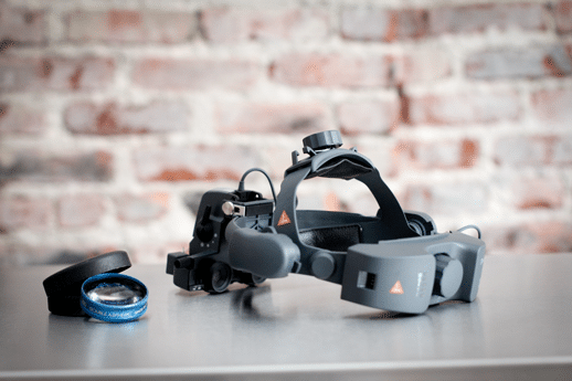
Indirect Ophthalmoscopy
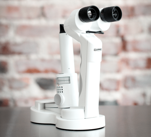
Slit-lamp
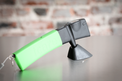
Tonometer : The Tonovet
EQUIPMENT FOR THE ADDITIONAL EXAMS (ERG)
ELECTRORETINOGRAM (ERG)
Your pet can be affected by a disease of the retina (nervous –sensitive part of the eye located at the far back) that can make your animal blind. The retina can be directly examined but its function can also be assessed with an additional test called electroretinogram that is done under sedation.
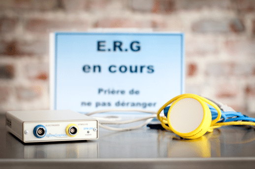
Electroretinogram
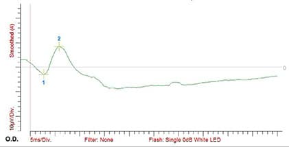
Graph of a normal retinal function
OCULAR ULTRASOUND MACHINE
An ocular ultrasound can be recommended when the intra-ocular structures are note directly visible or if there is a condition affecting the orbital space, behind the eye (inflammation, abscess, cancer) or during an assessment prior to cataract surgery.
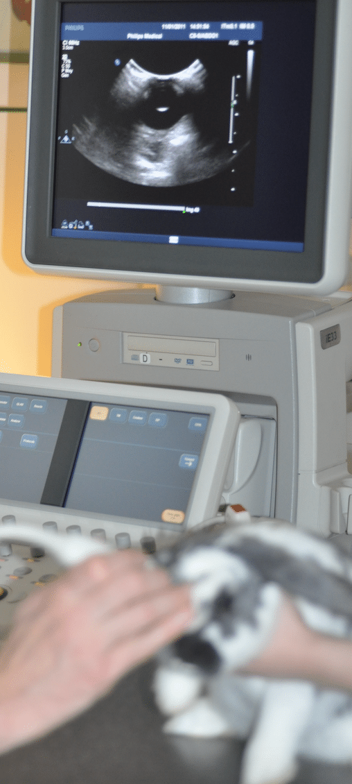
Ocular ultra-sound
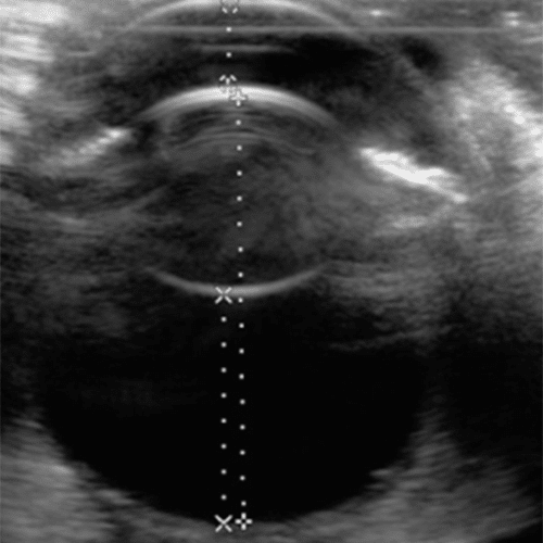
Image by ultrasound of a normal eye
EQUIPMENT USED DURING SURGERY FOR THE ANESTHESIA AND THE MONITORING OF THE ANIMAL DURING ANESTHESIA
All the ocular surgeries will need to be performed under anesthesia. Physical exam and blood work prior to surgery will therefore be necessary in order to check the health status of your pet and be sure that the risk due to anesthesia are minimal. These preoperative results will also allow us to establish the anesthesia protocol that is adequate for its condition
The most modern anesthesia system and a monitoring system checking various vital parameters will be used during the surgery. This equipment allow us to perform surgery in the most secure conditions possible.
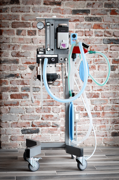
Anesthesia machine
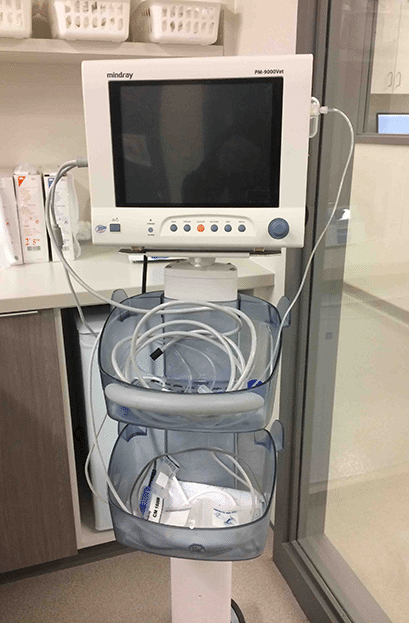
Monitoring machine used during anesthesia
MICROSURGERY INSTRUMENTS
Because of the small size of the lesions and the fragility of the ocular tissues on which surgeries are performed, very thin and delicate instruments are necessary for most of the ocular surgeries.
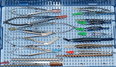
Microsurgical instruments
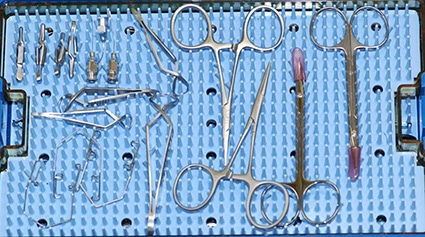
Microsurgical instruments
SURGICAL MICROSCOPE
During all surgeries on the cornea and in the eye (such as a cataract surgery), an operating microscope is necessary in order to have a very precise view of the tissues. An automated surgical microscope include light sources and various magnifying lenses allowing to reach a magnification of 20 times.
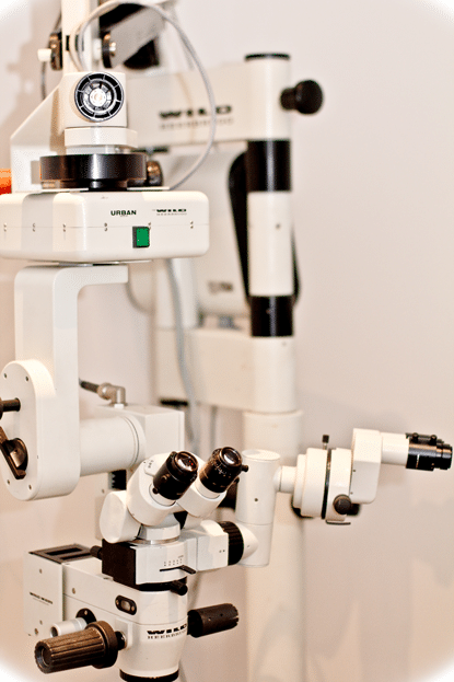
Microsurgical microscope
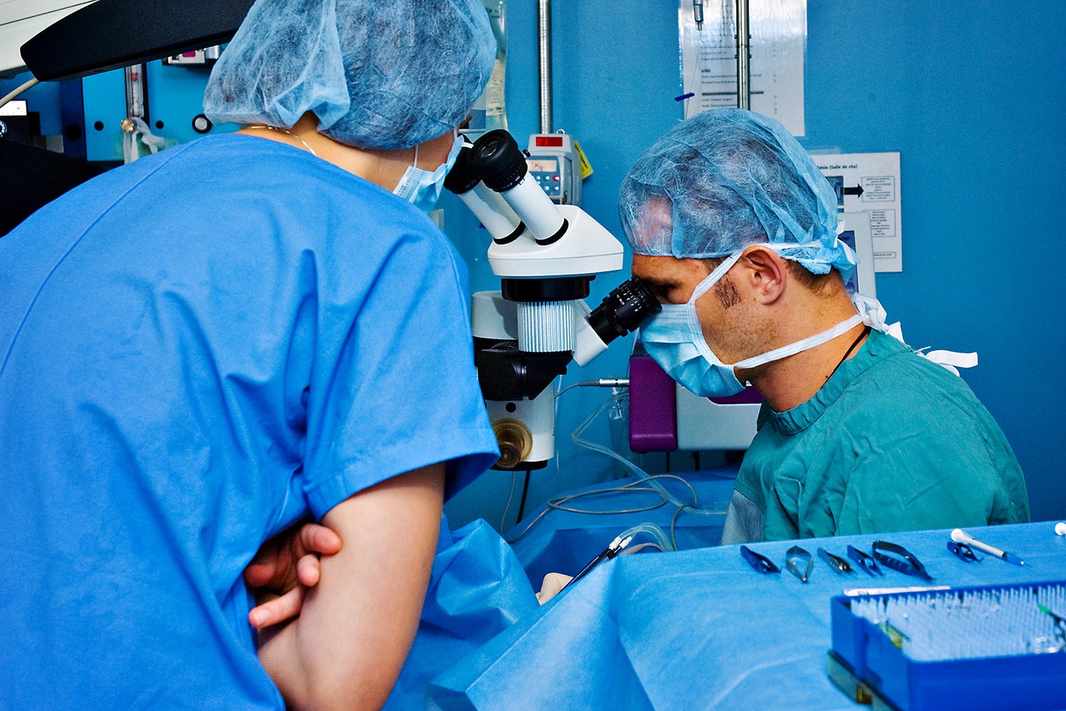
Microsurgical microscope
PHACOEMULSIFICATION MACHINE
There is no medical treatment to cure (remove) a cataract.
The surgical removal of a cataract (opacity of the lens that cause loss of vision) is the only possible treatment to give or improve the vision of your pet with cataract.
The surgical technique consists of a phaco-emulsification (removal of the opaque masses located in the bag of the lens) with the option to insert an artificial lens in the emptied lens bag. In order to perform this type of surgery, the animal needs to be anesthetized and we use the surgical microscope, the microsurgical instruments and the phacoemulsification machine. It is the same surgical procedure as when done for humans.
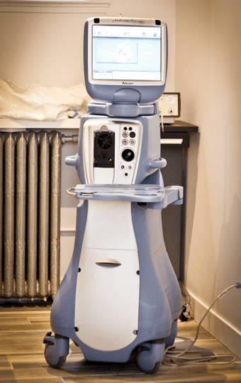
Phaco-emulsification machine
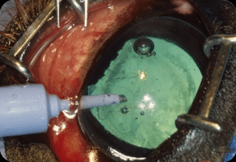
View of a cataract surgery by phaco-emulsification

