Some ocular diseases can progress very quickly and lead to the loss of the vision and/or the loss of the eye. This document reviews the signs that should be an alert for the pet’s owners.
Signs of ocular pain
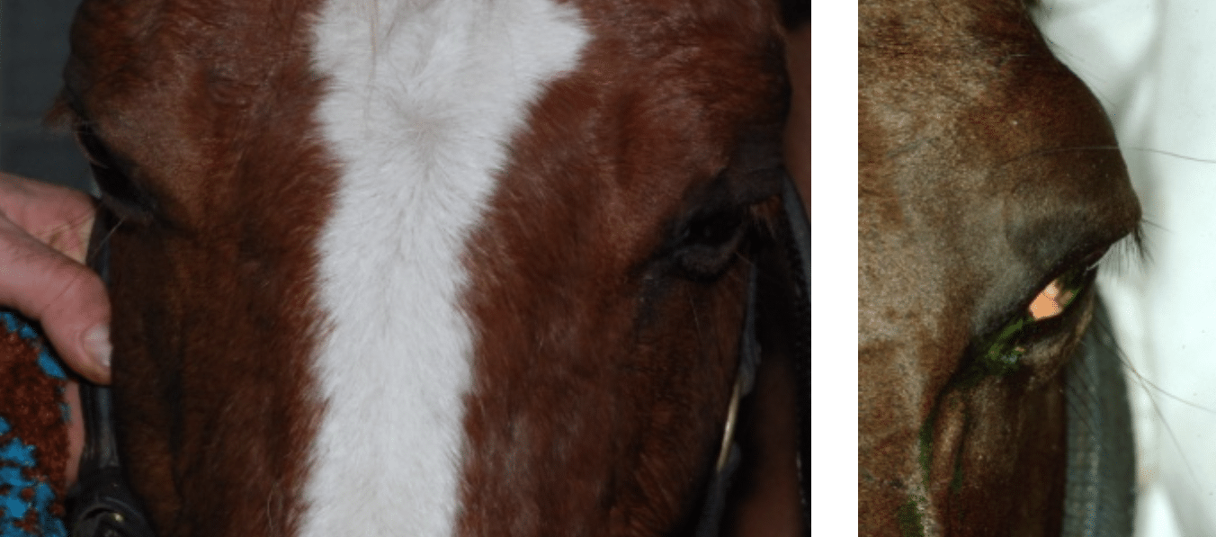
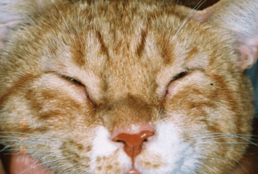
Blepharospasm/rubbing
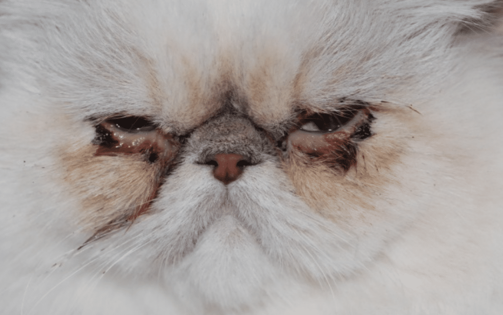
It is very frequent to observe blepharospasm when an animal has an ocular disease: he will blink the eyelids more often. He might keep his eye shot and might try to rub it to relieve the pain. However, few breeds (dogs and cats with round head and flat face such as Pugs, Pekinese, Bulldog, Shih Tzus, Persians) have a less sensitive cornea and do not blink that much.
Ocular discharge
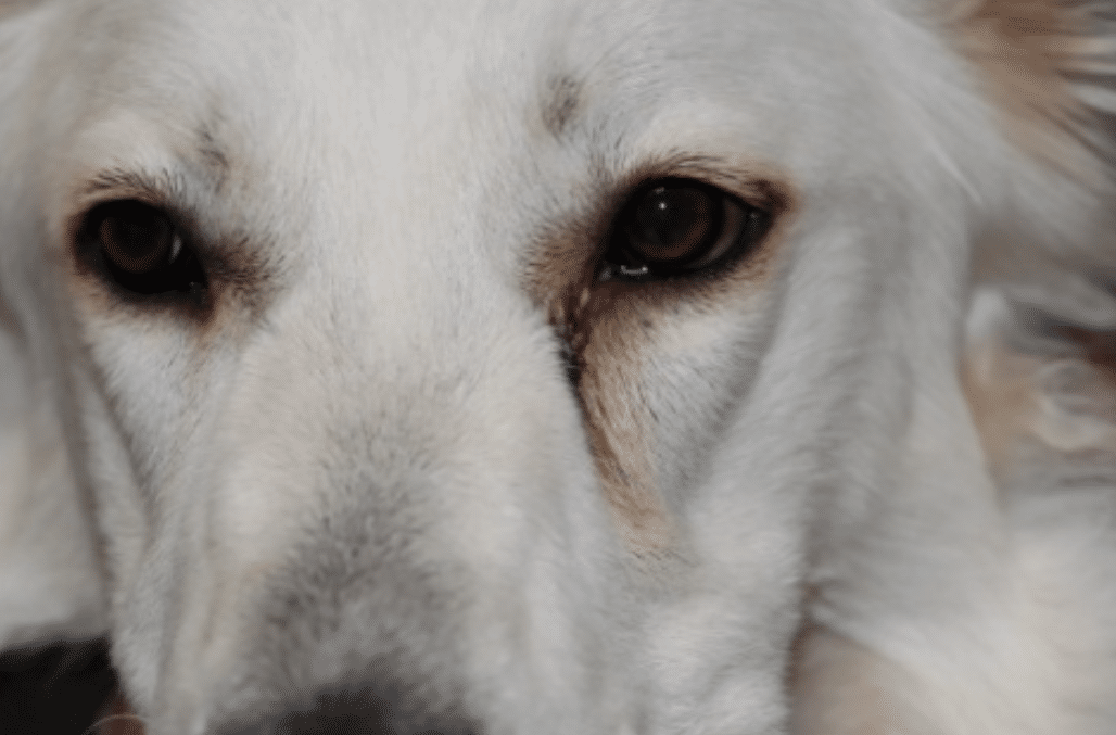
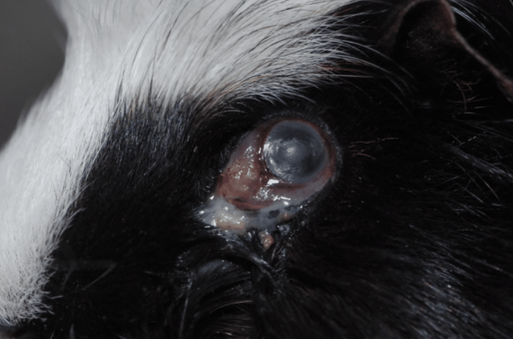
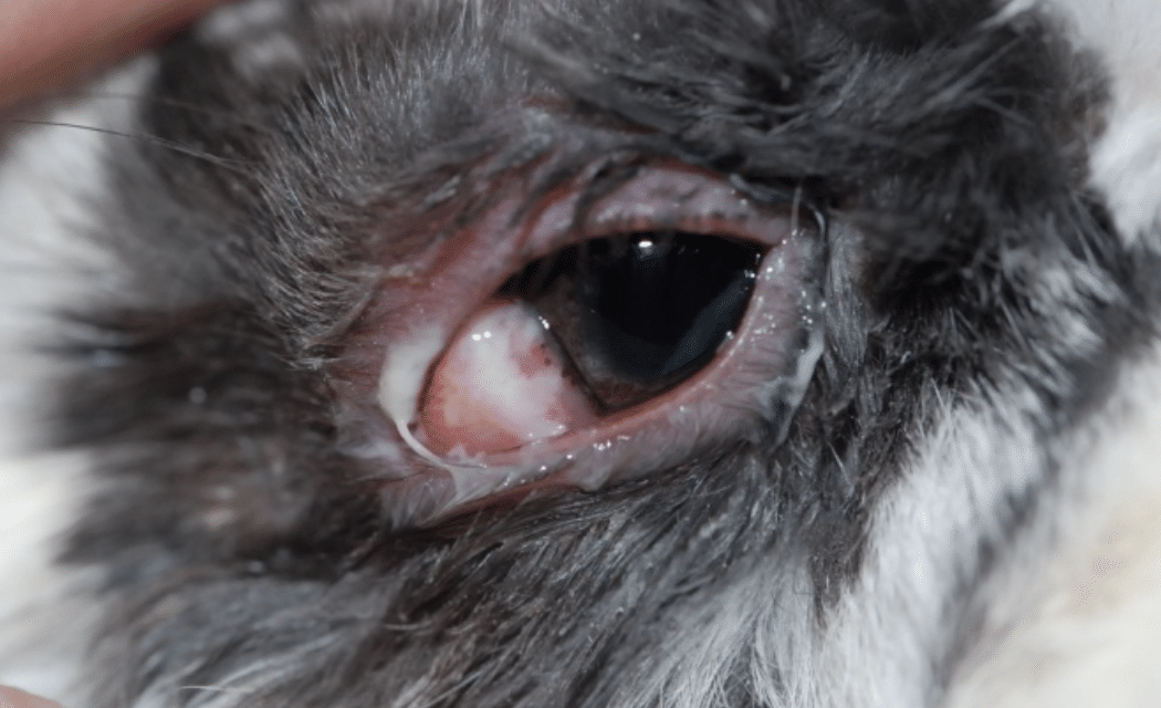
Very often, a discharge can be observed at the level of the eye and the nare. The discharge van be clear (aqueous), mucous, purulent, with a yellowish/greenish (if a bacterial infection is present) even brownish.
Change of the aspect of the eye
Position of the eye
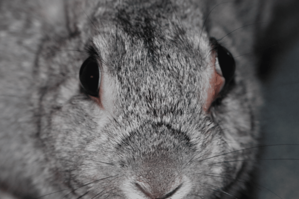
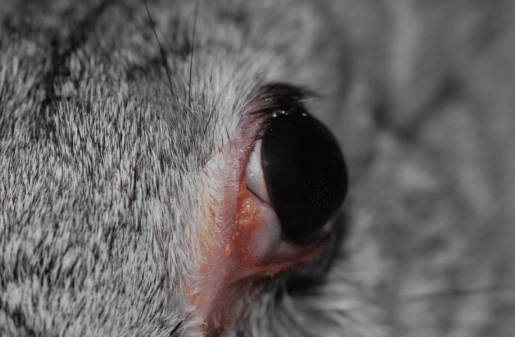
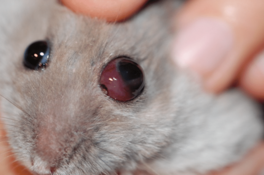
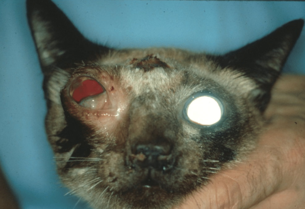
The position of the eye can be compared to the other eye. Sometimes, the eye can be more inside or enophtahlmia (the ocular pain can lead the eye to be inside as a mean to protect it) or more outside or exophthalmia. This can be due to a mass behind the eye (cancer or tooth abscess) that pushes the globe outside of the orbital cavity.
Redness of the conjunctiva
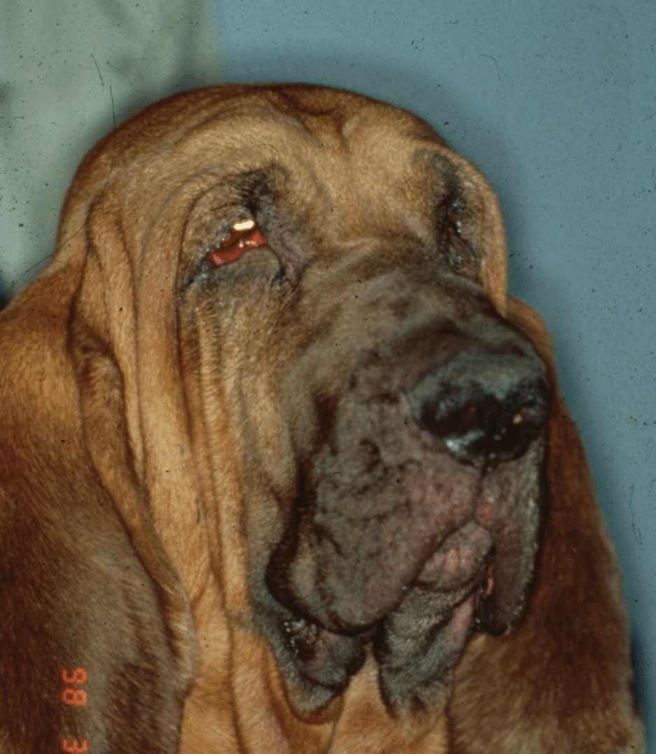
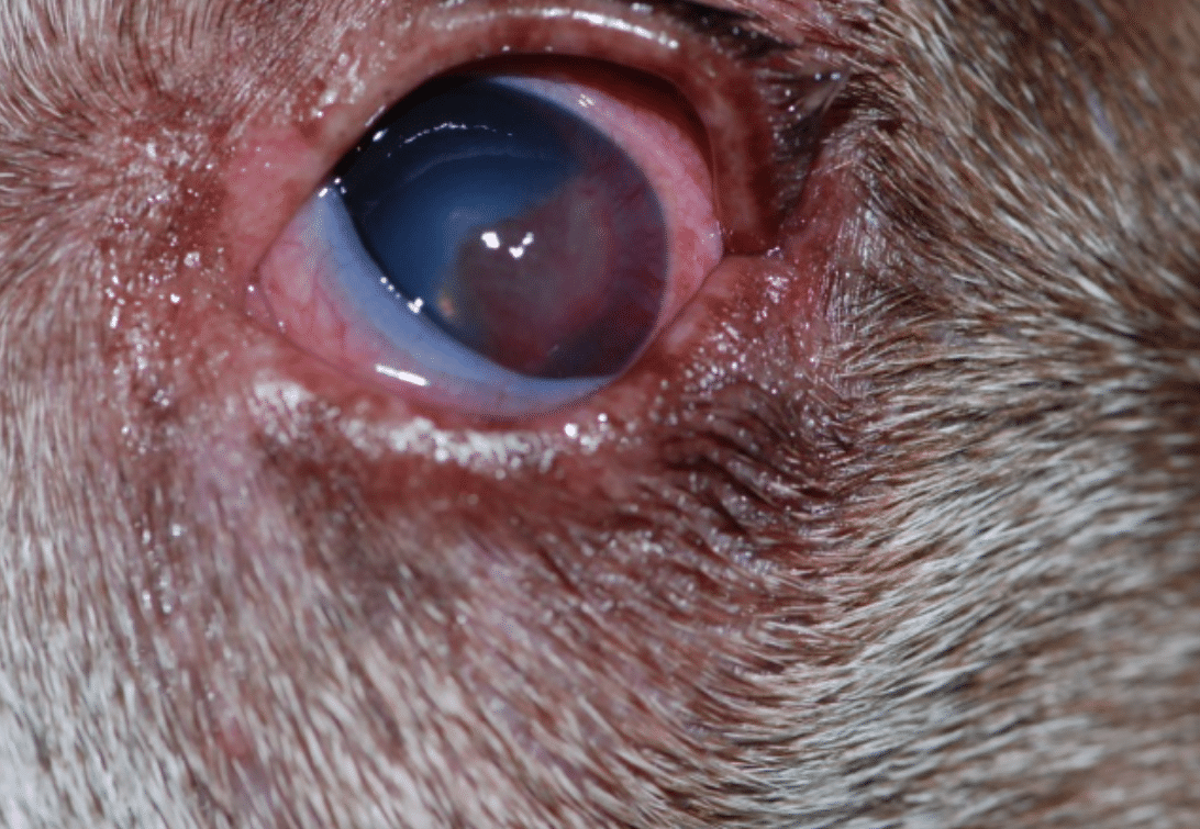
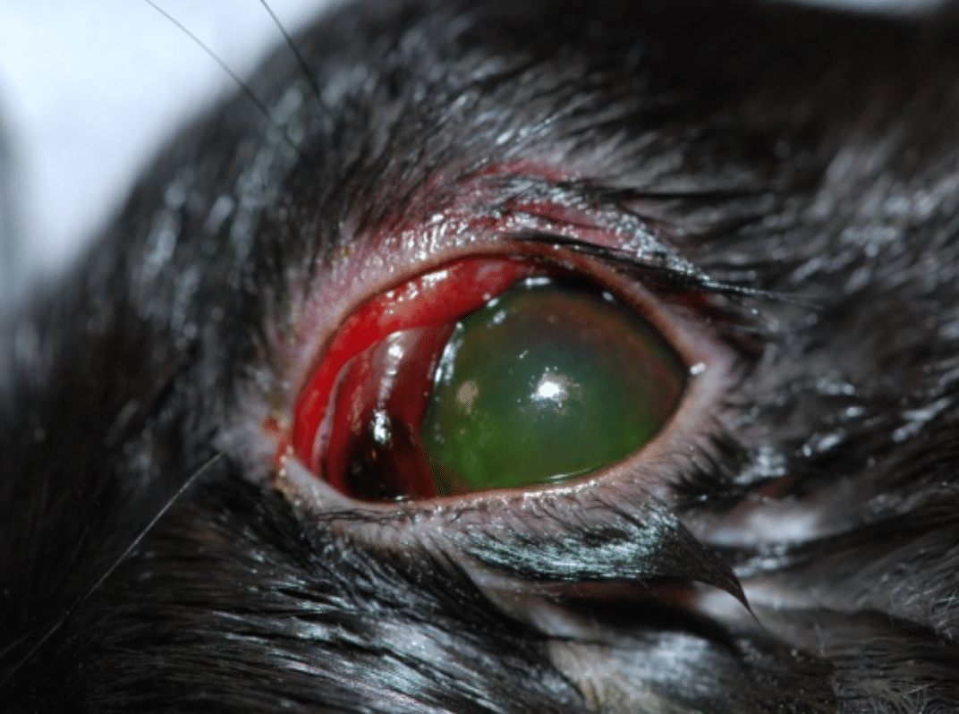
The redness of the conjunctiva is an ocular sign that is observed in many ocular diseases. They can be benign simple inflammation of the conjunctiva (or conjunctivitis) or severe (corneal ulcer, glaucoma). It is an ocular sign fairly easy to see but it does not indicate the severity of the ocular disease.
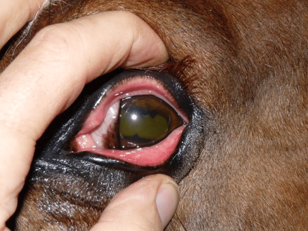
Redness of the conjunctiva in a horse (uveitis)
Redness of the conjunctiva in a dog (corneal ulcer)
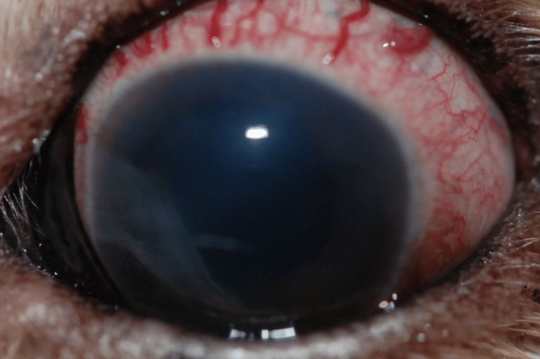
New tortuous vessels
In case of glaucoma (increase of the pressure in the eye), some vessels stretch and appear tortuous on the white part of the eye (the sclera). It can difficult to distinguish them from the vessels from the conjunctiva when this one is inflamed. However, these tortuous vessels are a sign of glaucoma. Ocular disease that needs to be treated as soon as possible as it leads to severe ocular pain and irreversible blindness very rapidly.
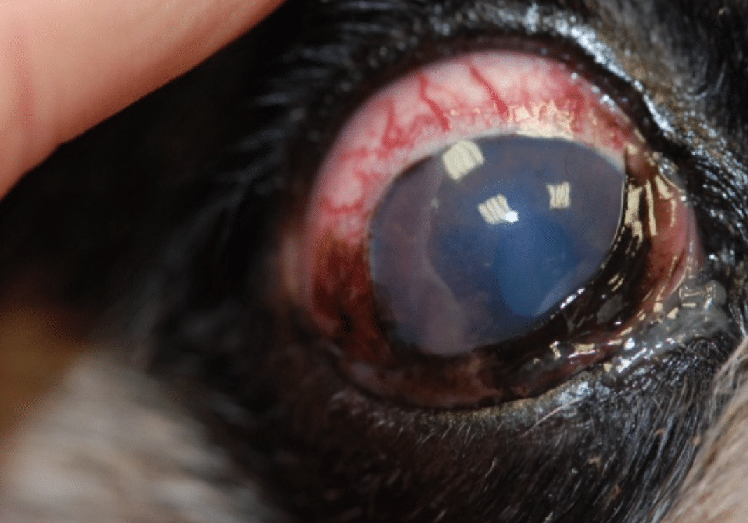
Velum on the cornea that is normally transparent
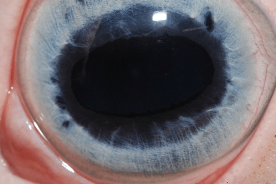
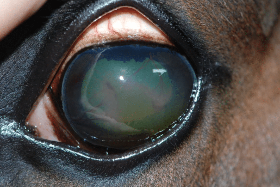
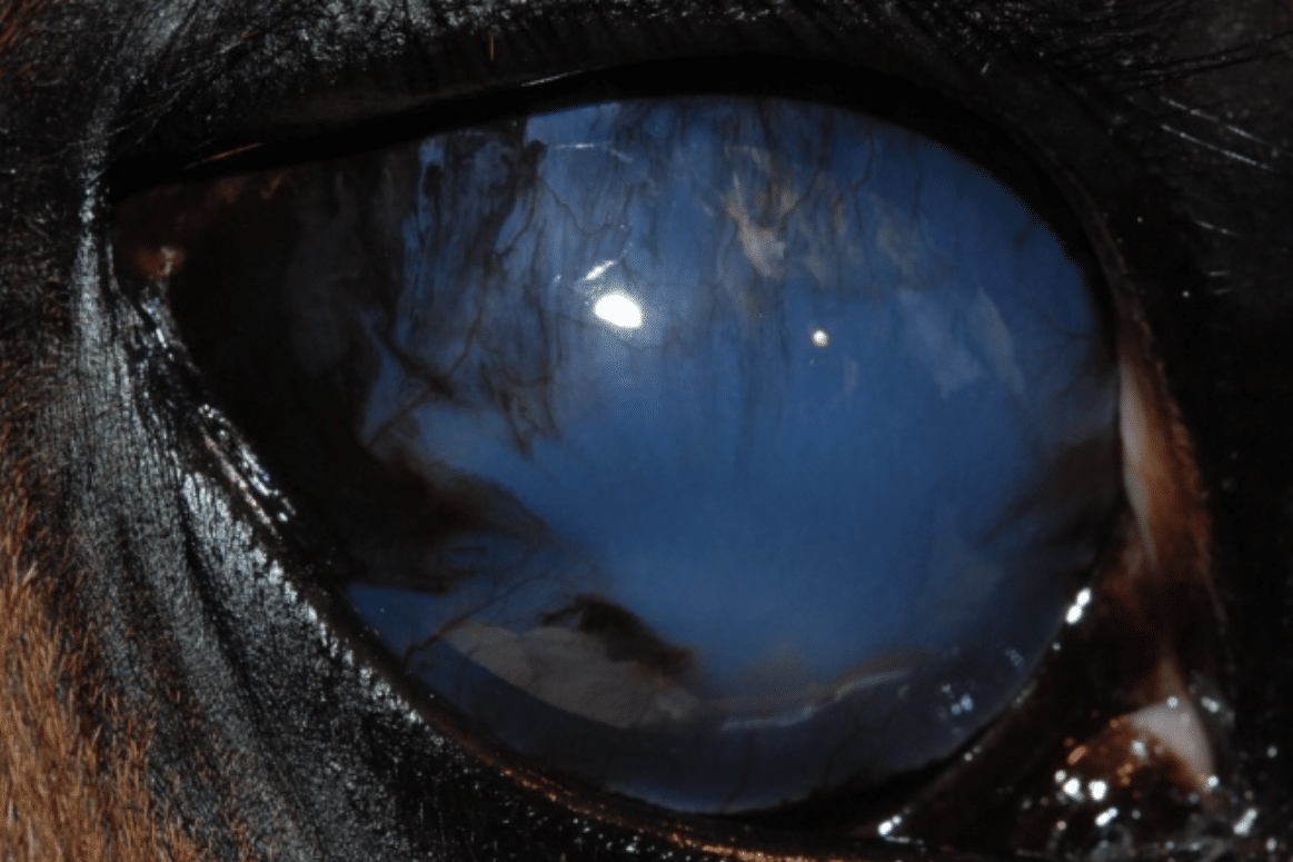
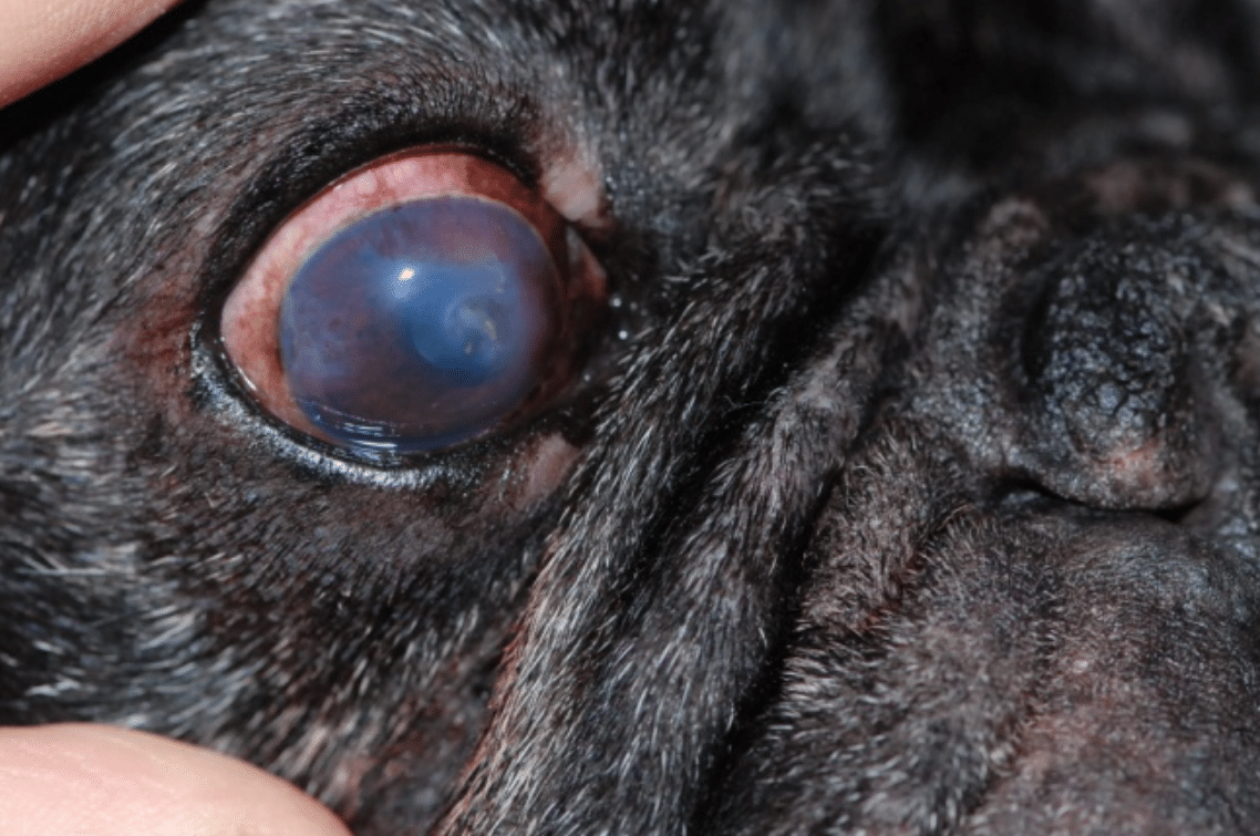
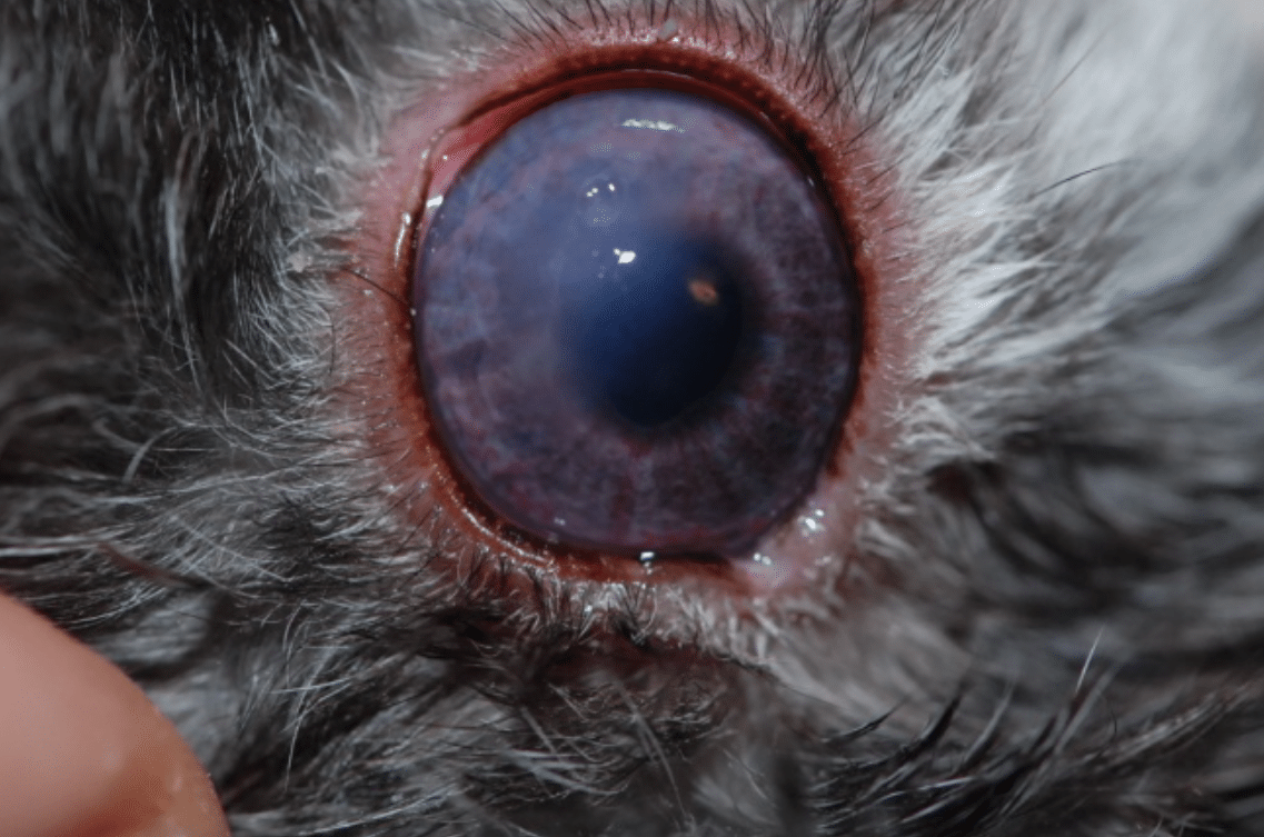
The cornea is the windshield of the eye the light and the image gets to the eye through the cornea. The cornea is completely transparent, without vessel, without pigment. A whitish, bluish “foggy” aspect of the cornea can be observed in case of corneal ulcer (corneal wound), uveitis (inflammation in the eye), glaucoma (increase of pressure in the eye). This ocular sign is usually a sign of a disease that needs to be treated rapidly, and therefore the owner should seek a veterinary advice if the sign is observed.
Changes in the behavior of the animal
Any unusual behavior should alert the owner, like:
It is very important that the owner observe unusual behavior of the pet as well as signs that are expression of ocular disease. We recommend to rapidly seeking the advice of your regular veterinarian who will establish a diagnosis, evaluate the severity of the disease, start a treatment and eventually refer you to a veterinary ophthalmologist.

